
Article Categories
- All Categories
-
 Data Structure
-
 Networking
-
 RDBMS
RDBMS
-
 Operating System
-
 Java
Java
-
 MS Excel
-
 iOS
iOS
-
 HTML
HTML
-
 CSS
CSS
-
 Android
-
 Python
-
 C Programming
-
 C++
C++
-
 C#
C#
-
 MongoDB
-
 MySQL
MySQL
-
 Javascript
-
 PHP
PHP
-
 Economics & Finance
Agarose Gel Electrophoresis: Definition, Principle, Steps and Uses
Introduction
Agarose gel electrophoresis is a laboratory technique used to separate DNA, RNA, or proteins based on their size and charge.
It is an essential method in molecular biology, biochemistry, and genetics research, and is widely used for the analysis of nucleic acids in forensic, medical, and biotechnological applications.
Definition of Agarose Gel Electrophoresis
Agarose gel electrophoresis is a technique that involves the separation of nucleic acids or proteins in a gel matrix made of agarose.
The matrix is placed in an electric field, and the charged molecules migrate through the matrix based on their size, shape, and charge.
Smaller molecules migrate faster than larger ones, leading to the separation of the sample into distinct bands that can be visualized and analysed.
Principle of Agarose Gel Electrophoresis
The principle of agarose gel electrophoresis is based on the fact that DNA, RNA, and proteins are negatively charged molecules that can be separated based on their size and charge by applying an electric field.
The gel matrix used in agarose gel electrophoresis is made of agarose, a polysaccharide extracted from seaweed that forms a porous structure when mixed with water.
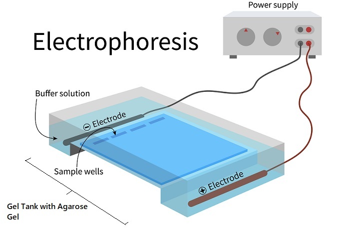
The agarose gel acts as a sieve, allowing smaller molecules to pass through more easily than larger ones. The electric field is applied by placing the gel in a buffer solution and applying an electrical current across the gel using two electrodes.
The DNA, RNA, or protein sample is mixed with a loading buffer, which contains a tracking dye that allows the progress of the electrophoresis to be monitored.
The sample is loaded onto the gel, and the electric current is turned on. The negatively charged molecules in the sample migrate through the gel towards the positively charged electrode
The smaller molecules migrate more quickly through the gel and reach the end of the gel first, while the larger molecules take longer to migrate and form distinct bands on the gel. The bands can be visualized using a staining agent, such as ethidium bromide, which binds to the nucleic acid molecules and fluoresces under UV light.
Steps of Agarose Gel Electrophoresis
The following are the steps involved in agarose gel electrophoresis ?
Preparation of The Gel
The first step in agarose gel electrophoresis is the preparation of the gel. Agarose is mixed with a buffer solution, such as Tris-borate-EDTA (TBE) or Tris-acetate-EDTA (TAE), to create a gel matrix.
The concentration of the agarose gel can vary depending on the size range of the molecules being analysed. Higher concentrations of agarose are used for the separation of smaller molecules, while lower concentrations are used for larger molecules.
The gel matrix is poured into a casting tray and allowed to solidify. Once the gel has solidified, it is removed from the casting tray and placed in a gel electrophoresis chamber.
Preparation of The Sample
The sample to be analysed is prepared by mixing it with a loading buffer, which contains a tracking dye that allows the progress of the electrophoresis to be monitored.
The loading buffer also contains a denaturant, such as sodium dodecyl sulphate (SDS), which denatures the protein and gives it a negative charge.
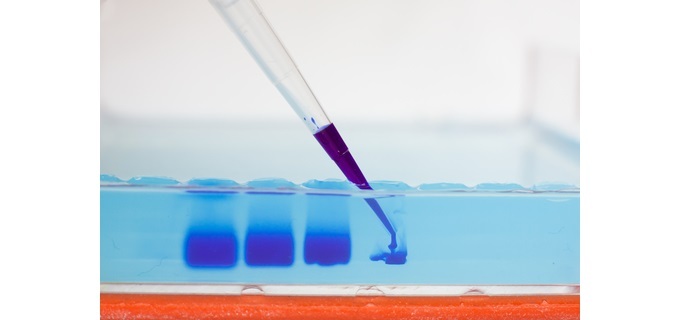
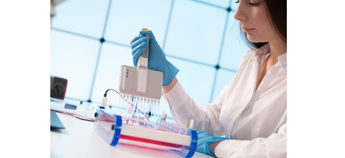
The sample is loaded into wells at one end of the gel using a micropipette. Care should be taken not to overload the wells, as this can lead to distortion of the bands.
Running The Gel
Once the sample has been loaded onto the gel, the electric current is turned on, and the gel is allowed to run for a specific period of time.
The length of time required for the electrophoresis run depends on the size range of the molecules being separated and the concentration of the agarose gel. The gel is typically run until the tracking dye reaches the end of the gel.
Staining and Visualization
Once the electrophoresis run is complete, the gel is stained with a suitable staining agent to visualize the separated molecules.
Ethidium bromide is a commonly used staining agent for nucleic acids, while Coomassie Blue is used for proteins.
The gel is illuminated with UV light to excite the staining agent, and the separated molecules appear as distinct bands on the gel. The bands can be photographed or analysed using densitometry to determine their size and quantity.
Uses of Agarose Gel Electrophoresis
Some of the most common uses of agarose gel electrophoresis are ?
DNA and RNA Analysis
Agarose gel electrophoresis is widely used for the analysis of DNA and RNA in various applications such as PCR, DNA sequencing, and RNA isolation. The technique can be used to separate DNA fragments based on their size, which is essential for downstream applications such as cloning and gene expression analysis.

Protein Analysis
Agarose gel electrophoresis is also used for the separation and analysis of proteins. The technique is particularly useful for separating proteins based on their size and charge, which can provide insights into their structure and function.
Forensic Analysis
Agarose gel electrophoresis is used in forensic analysis to identify DNA samples from crime scenes. The technique is used to separate DNA fragments from different individuals and compare them to the DNA sample collected from the crime scene.
Medical Diagnosis
Agarose gel electrophoresis is used in medical diagnosis to identify genetic mutations associated with various diseases such as sickle cell anaemia and cystic fibrosis. The technique is used to separate and analyse DNA fragments associated with the disease, which can help in diagnosis and treatment.
Conclusion
Agarose gel electrophoresis is a fundamental technique in molecular biology, biochemistry, and genetics research. The technique allows the separation and analysis of nucleic acids and proteins based on their size and charge, providing valuable insights into their structure and function.
Agarose gel electrophoresis is a versatile technique used in various applications such as PCR, DNA sequencing, forensic analysis, and medical diagnosis. It is a simple and cost-effective technique that has become an essential tool in molecular biology and genetics research.

18K+ Views
